
What does mindfulness actually do to your brain? I decided to start off with the most concrete examples. Literally, those who meditate and those who don’t have different looking (and measurably functioning) brains, a bit like the people who go to the gym tend to look different to who don’t.
It’s understood at this point that it leads to structural change. In short, it changes the connections between different neurons (brain cells). It strengthens the pathways that are helpful to mindfulness and weakens ones that interfere with it. This is also known as neuroplasticity: the changing of neurons, to put it literally. This is a bit too high level for some people so I wanted to delve a little deeper.
Functional magnetic resonance imaging, fMRI, studies are of questionable significance, but then again, everything is. Many scientists hypothesised whether there is a change in the connectivity between default mode network (DMN) regions.
A DMN is basically a collection of parts of the brain that like to be in sync with each other when we are at rest. These parts are especially active when we’re not involved in a goal-directed task, e.g. when we are day dreaming. Sometimes the DMN gets shut off when we are actually doing something goal-directed. It can be hard to imagine what a brain network is without naming a specific part of the brain. The difficulty with naming it… Is evident when you try. But we shall try. What has been conventionally included in the DMN is as follows: posterior cingulate cortex, precuneus, medial prefrontal cortex, angular gyrus, dorsomedial prefrontal cortex… The list goes on. It is mostly a variety of cortical regions. Interestingly, it overlaps with parts of the limbic system at the hippocampus.
So what fMRI studies found was that those who practice meditation had weaker functional connections between certain DMN regions – especially those associated with emotional judgement. On the flipside, meditators had stronger links in other regions (specifically the dorsomedial prefrontal cortex and right inferior parietal lobule). It is difficult to articulate what exactly this means in practical terms, but it’s real – whatever it is. You can read more about these studies here and here.
Mindfulness is associated with greater attention related activity in the anterior dysgranular insula regions. This suggests that mindfulness is linked to being able to find better context for internal sensations given one’s the surroundings. In other words, it helps to answer the question – why do I feel a this way in these circumstances? (See study) On a related note, another study suggested that this ellusive non-reactivity that we are all after is inversely related with insula activation. Mindfulness could reduce vulnerability to depression by reducing automatic emotional responding via the insula.
A different type of MRI, so called diffusion tensor imaging, or DW-MRI, has also been used to study those who practice meditation.
Interestingly, those who practice mindfulness have greater cortical thickness in the anterior regions of the brain: the medial prefrontal cortex, superior frontal cortex, temporal pole and the middle and interior temporal cortices. On the flipside, there was reduced cortical thickness in the posterior regions of the brain including the postcentral cortex, inferior parietal cortex, middle occipital cortex and posterior cingulate cortex. These and other findings indicate that those that take part in meditation over long periods have structurally different brains in terms of both gray and white matter. You can read more here: Lazar et al, Tang et al, Holzel et al, Luders et al, Kang et al.
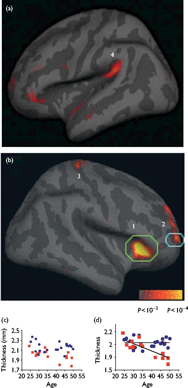
Image from Meditation experience is associated with increased cortical thickness showing the cortical regions that are thicker in meditators. Numbered regions: (1) insula, (2) Brodmann area (BA) 9/10, (3) somatosensory cortex, (4) auditory cortex.
Loving-kindness meditation has been linked to increased gray matter volume in the right angular and posterior parahippocampal gyri. The right angular gyrus has been linked to empathy, anxiety and mood.
Even if fMRI is just a mirage, DW-MRI seems to back up the findings of changing structure. It is fascinating to think that we can actively structurally change our brains.
Besides the different types of MRI, changes have been shown on EEG (it’s like an ECG/EKG for the brain). More importantly, those who do mindfulness tend to behave differently – as shown by their differing test scores in various tasks – I shall describe this another time.

Very interesting, though I am no scientist. It was easy to follow, and I certainly learned something.
LikeLiked by 1 person
Glad you did, means a lot, thanks for the comment
LikeLiked by 1 person